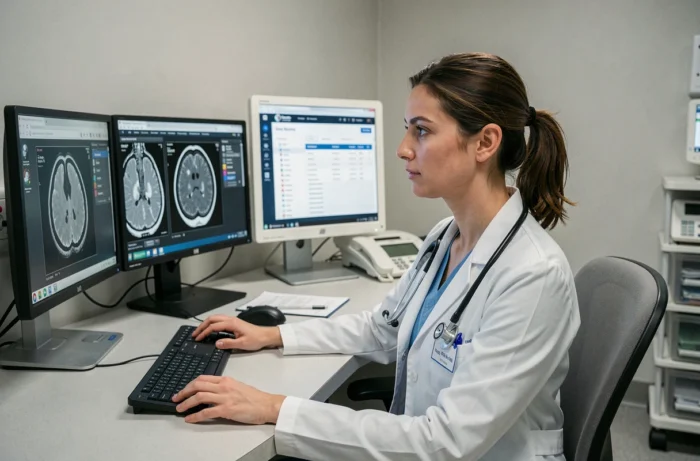
When you undergo a medical imaging exam (MRI, CT scan, X-ray), you often leave with a paper report and sometimes a CD-ROM. The actual journey of these images between the radiology office, the primary care physician, and the specialist remains unclear for most patients. Understanding how your images circulate, where they are stored, and who can access them changes the way you manage your medical follow-up.
What happens technically between the acquisition and consultation of your images
At the moment the radiology technician initiates the acquisition, the images are generated in DICOM format, the international standard used by all imaging devices. These files are large: a single series of MRI can represent several hundred slices.
The images then transit to a system called PACS (Picture Archiving and Communication System). It is the central server that stores, archives, and redistributes your images. The radiologist accesses it from their console to interpret the exam, write the report, and validate the results.
Meanwhile, a RIS (Radiology Information System) software manages all the administrative aspects: patient identity, type of exam, appointment date, history. It is the link between your file and your images. When these two components work together, it becomes clearer how the web service irld.net for medical images allows tracking this journey from a simple browser.
The point that often escapes patients: your images do not remain on the examination device. They migrate to secure servers, sometimes shared among several offices in the same network. In Rennes, for example, the GIE IRLDR connects several dozen private radiologists to a common IT tool (RIS-PACS ONE MANAGER), allowing a radiologist to consult your previous images even if they were taken in another office in the network.
Distribution of imaging results: web portal versus CD-ROM
The CD-ROM burned after the exam has long been the only means of distribution. It poses several practical problems: it can get scratched, CD drives are disappearing from recent laptops, and it does not allow for any updates if the radiologist completes their interpretation after your departure.
Web portals for image distribution have replaced this medium in most structured imaging centers. The principle is simple:
- After the report is validated, the office makes your images available on a secure space accessible via a link and a personal code.
- Your primary care physician receives a notification (often via secure health messaging) and can directly view the images on their screen, without needing to install any software.
- You retain patient access to download your images or share them with a specialist for a second opinion.
Access to images no longer depends on a physical medium. The general practitioner in Cesson-Sévigné or Saint-Grégoire consults the same images as the surgeon in Rennes, with the same diagnostic quality. Feedback varies on the speed of online availability depending on the centers, but the delay is generally counted in hours after the report is validated.
What the patient can check
By logging into their results space, one can verify that all series of images are present. An MRI exam often includes several sequences (T1, T2, diffusion). If a series is missing, it is better to contact the office’s secretariat before the follow-up appointment.
Security and hosting of medical imaging data in France
Medical images are personal health data. Their hosting is subject to strict requirements. In France, any health data host must be certified HDS (Health Data Host), a certification granted after an audit by an accredited organization.
A French decree from March 2026 now requires a certificate of compliance for any digital health service, issued by the National Health Digital Agency (ANS). This certificate covers security, hosting, and interoperability. A graduated sanctions regime accompanies this obligation, changing the game for image distribution platforms.
At the European level, the regulation on the European Health Data Space (EHDS, EU Regulation 2025/327) came into effect on March 26, 2025. It makes the interoperability of medical records legally enforceable, with a gradual implementation. A specific extension for medical imaging is expected around 2031.
In practical terms, this means that platforms distributing your images must ensure that these images can be read and utilized by any compliant system, even in another European country. For a patient who consults in Rennes and then has surgery in Barcelona, this is a major change.
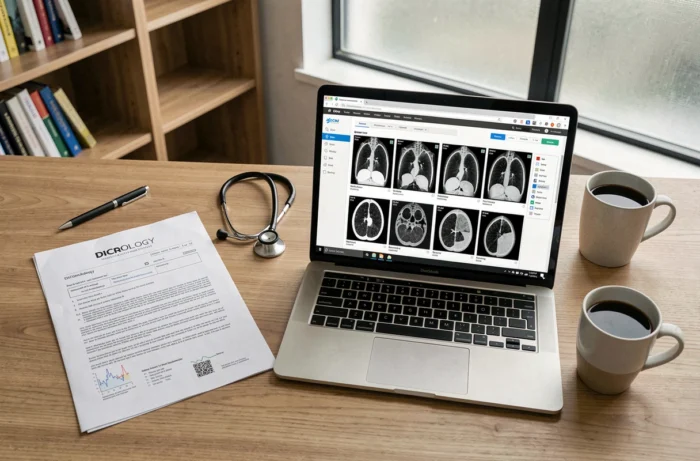
Medical imaging and My Health Space: what works today
My Health Space (formerly DMP) was supposed to centralize the entire medical file, including imaging. In practice, the feeding of the DMP with images remains partial. Textual reports are indeed uploaded, but large DICOM files still pose technical integration issues.
The Ségur digital health initiative, with its wave 2, has pushed software publishers to have their solutions referenced for secure data sharing. Several EHR (Electronic Health Record) publishers have obtained this referencing, which streamlines the transmission chain between the hospital, the city office, and the patient space.
For imaging exams performed in a center in Rennes or its outskirts (Cesson-Sévigné, Saint-Grégoire), the most reliable circuit today remains the combination of a dedicated distribution portal and secure health messaging. My Health Space will complement this system as the interoperability constraints of the EHDS regulation come into effect.
The journey of your medical images relies on a technical infrastructure that is rapidly evolving under regulatory pressure. Checking that your examination center offers secure web access to your results remains the most useful action to maintain control over your imaging file.